INTRODUCTION
"Micro" refers to tiny, "scope" refers to view or look at. Microscopes are tools used to enlarge images of small objects that can not be seen by naked eye so as they can be studied.
There are many type of microscopes such as electron microscope and ultramicroscope but the most widely used are the optical microscope or the light microscope. The light microscope is an instrument which uses two lenses which magnifies and helps to focus the image of the small object.

Part of microscope
|
Function
|
Eyepiece Lens
|
the lens at the top that you look through. They are usually 10X or 15X power.
|
Tube
|
Connects the eyepiece to the objective lenses
|
Arm
|
Supports the tube and connects it to the base
|
Base
|
The bottom of the microscope, used for support
|
Illuminator
|
A steady light source (110 volts) used in place of a mirror. If the microscope has a mirror, it is used to reflect light from an external light source up through the bottom of the stage.
|
Stage
|
The flat platform where the slides is placed.
Stage clips hold the slides in place.
Mechanical stage able to move the slide around by turning two knobs. One moves it left and right, the other moves it up and down.
|
Revolving Nosepiece or Turret
|
This is the part that holds two or more objective lenses and can be rotated to easily change power.
|
Objective Lenses
|
There are 4 objective lenses on a microscope. They almost always consist of 4X, 10X, 40X and 100X powers. When coupled with a 10X (most common) eyepiece lens, we get total magnifications of 40X (4X times 10X), 100X , 400X and 1000X. To have good resolution at 1000X, you will need a relatively sophisticated microscope with an Abbe condenser. The shortest lens is the lowest power, the longest one is the lens with the greatest power. Lenses are color coded and if built to DIN standards are interchangeable between microscopes. The high power objective lenses are retractable (i.e. 40XR). This means that if they hit a slide, the end of the lens will push in (spring loaded) thereby protecting the lens and the slide. All quality microscopes have achromatic, parcentered, parfocal lenses.
|
Rack Stop
|
This is an adjustment that determines how close the objective lens can get to the slide. It is set at the factory and keeps students from cranking the high power objective lens down into the slide and breaking things. Adjust this if very thin slides been using and the specimen unable to focus on high power.
|
Condenser Lens
|
The purpose of the condenser lens is to focus the light onto the specimen. Condenser lenses are most useful at the highest powers (400X and above). Microscopes with in stage condenser lenses render a sharper image than those with no lens (at 400X).
|
Diaphragm or Iris
|
Many microscopes have a rotating disk under the stage. This diaphragm has different sized holes and is used to vary the intensity and size of the cone of light that is projected upward into the slide. There is no set rule regarding which setting to use for a particular power. Rather, the setting is a function of the transparency of the specimen, the degree of contrast you desire and the particular objective lens in use.
|
Magnification and Resolution
| |||||||||||||||||
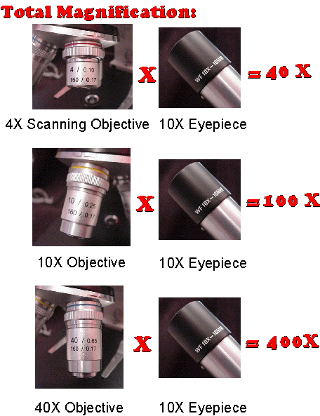
| Total Magnification: | |||||||||||||||||
| To figure the total magnification of an image that you are viewing through the microscope is really quite simple. To get the total magnification take the power of the objective (4X, 10X, 40x) and multiply by the power of the eyepiece, usually 10X.Total magnification = Objective lens power x eyepiece lens power | |||||||||||||||||
 OBJECTIVE
-To learn the importance of magnification and resolution of microscope.
-To learn the ways to take care of the microscope.
MATERIALS AND REAGENT
Microscope slide and cover-slip.
PROCEDURE
(refer to manual)
RESULT

-We observed that magnification enebles us to see speciment in details, while resolution enebles us to see clearer object. We find that both magnification and resolution is important for us to get a quality image of speciment.
-Thus, microscope is an expensive instrument.
REFERENCES http://www.microscope-microscope.org/basic/microscope-parts.htm http://en.wikipedia.org/wiki/Microscope 1.2 EXAMINATION OF CELL INTRODUCTION Historically, the study of cell biology could not have happened without the invention of microscopes because cells were not known to exist before Antonin van Leeuwenhoek and Robert Hooke saw them in their primitive microscopes Today, much cell biology research still requires careful microscopic examination of cells and their internal structures. It is not too strong a statement to say that microscopy is the single most important tool for the cell biologist. OBJECTIVE To provide an experience in the use of microscope To illustrate the diversity of cell and microorganisms. MATERIAL AND REAGENTS -Culture -Immersion Oil -Lens Tissue -A Microscope slide containing stained microorganism -Inoculating loop -Bunsen Burner -Slide and coverslip PROCEDURE (refer to lab manual ) RESULT
DISCUSION
Lactobacillus
Lactobacillus are
rod-shaped, Gram-positive, fermentative, organotrophs. They are usually
straight, although they can form spiral or coccobacillary forms under certain
conditions. They are often found in pairs or chains of varying length.
Lactobacillus are classified as lactic acid bacteria, and derive almost all of
their energy from the conversion of glucose to lactate during homolactic
fermentation. In this process 85-90% of the sugar utilized is converted to
lactic acid. They generate ATP by nonoxidative substrate-level phosphorylation.
Gram positive bacteria
In general, the following characteristics are present
in .Gram-positive bacteria has the following characteristic, that is it they
have cytoplasmic lipid membrane, thick peptidoglycan layer, and they are also a
class of bacteria that take up the crystal
violet stain.
used in the Gram
staining method
of bacterial differentiation. The thick peptidoglycan layer in the cell
wall that
encases their cell
membrane retains
the stain, making definitive identification possible. Only some species have a capsule usually consisting of polysaccharides. Also only some species are flagellated, and when they are, they only have two rings to
support them.
CONCLUSION
Based on the experiment that we observe, The
Lactobacillus is a rod shape-bacteria. We found that some of them are straight
while others are spiral.
REFERENCE
| |||||||||||||||||


No comments:
Post a Comment