Sunday, 14 December 2014

LAB 5 :DETERMINATION OF ANTIMICROBIAL EFFECTS OF MICROBIAL EXTRACTS

Introduction :
Bacteriocins are an abundant and diverse group of ribosomally synthesized antimicrobial peptides produced by bacteria and archaea. Traditionally, bacteriocin production has been considered an important trait in the selection of probiotic strains, but until recently, few studies have definitively demonstrated the impact of bacteriocin production on the ability of a strain to compete within complex microbial communities and/or positively influence the health of the host. Although research in this area is still in its infancy, there is intriguing evidence to suggest that bacteriocins may function in a number of ways within the gastrointestinal tract. Bacteriocins may facilitate the introduction of a producer into an established niche, directly inhibit the invasion of competing strains or pathogens, or modulate the composition of the microbiota and influence the host immune system. Here we review the role of bacteriocin production in complex microbial communities and their potential to enhance human health.
During the last few years, a large number of new bacteriocins produced by lactic acid bacteria (LAB) have been identified and characterized. LAB-bacteriocins comprise a heterogeneous group of physicochemically diverse ribosomally-synthesized peptides or proteins showing a narrow or broad antimicrobial activity spectrum against Gram-positive bacteria. Bacteriocins are classified into separate groups such as the lantibiotics (Class I); the small (<10 kDa) heat-stable postranslationally unmodified non-lantibiotics (Class II), further subdivided in the pediocin-like and anti Listeria bacteriocins (subclass IIa), the two-peptide bacteriocins (subclass IIb), and the sec-dependent bacteriocins (subclass IIc); and the large (>30 kDa) heat-labile non-lantibiotics (Class III). Most bacteriocins characterized to date belong to Class II and are synthesized as precursor peptides (preprobacteriocins) containing an N-terminal double-glycine leader peptide, which is cleaved off concomitantly with externalization of biologically active bacteriocins by a dedicated ABC-transporter and its accessory protein. However, the recently identified sec-dependent bacteriocins contain an N-terminal signal peptide that directs bacteriocin secretion through the general secretory pathway (GSP). Most LAB-bacteriocins act on sensitive cells by destabilization and permeabilization of the cytoplasmic membrane through the formation of transitory poration complexes or ionic channels that cause the reduction or dissipation of the proton motive force (PMF). Bacteriocin producing LAB strains protect themselves against the toxicity of their own bacteriocins by the expression of a specific immunity protein which is generally encoded in the bacteriocin operon. Bacteriocin production in LAB is frequently regulated by a three-component signal transduction system consisting of an induction factor (IF), and histidine protein kinase (HPK) and a response regulator (RR). This paper presents an updated review on the general knowledge about physicochemical properties, molecular mode of action, biosynthesis, regulation and genetics of LAB-bacteriocins.


Objective:
To determine the antimicrobial effects of extracellular extracts of selected LAB strains.

Materials and reagents:
MRS broth
Sterile filter paper disk(50mm x 50mm)
Sterile forceps
Cultures of LAB and spoilage/phatogenic organism
Bench-top refrigenerated centrifuge
Incubator 30 celcius and 37 celcius
UV/V is spectrophotometer
Distilled deionized water
Trypticase soy agar
Brain heart infusion agar
Yeast extract


Procedure:

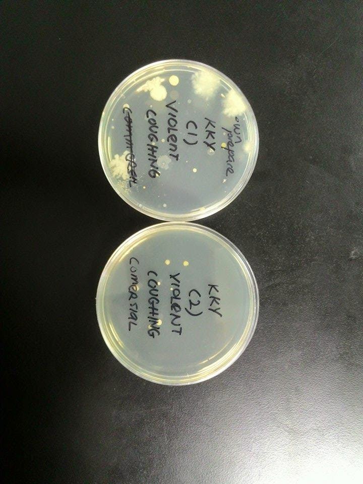
part 1:determination of bacteriocin activity via agar diffusion test
(refer to lab manual)
part 2:determination of bacteriocin activity via optical density
(refer to lab manual)

Result:

Strains of LAB
Strains of spoilage/ phatogenic bacteria
Inhibition Zone(cm)
1
E.coli
0.9
Staphylococcus aureus
0.8
2
E.coli
0.8
Staphylococcus aureus
0.7
avarage
E.coli
0.7
Staphylococcus aureus
0.8


Discussion:
Part 1 (Agar diffusion)
1) There is a wide number of bacteriocins produced by different Lactic Acid Bacteria.Bacteriocins are proteinaceous toxins produced by bacteria to inhibit the growth of similar or closely related bacterial strain(s). They are typically considered to be narrow spectrum antibiotics and are phenomenologically analogous to yeast and paramecium killing factors, and are structurally, functionally, and ecologically diverse.
2)Antimicrobial effect of LAB is mainly due to lactic acid and other organic acid production.    antimicrobial proteins provide an additional hurdle for spoilage and pathogenic microorganisms.
3)Staphylococcus Aureus is a gram positive coccal bacterium. Although its has thick cell wall made of polysaccharides and proteins but is easily digested by acid particularly acetic acid produced by lactic acid bacteria(LAB).
4)Escherichia coli is a gram negative bacteria .The cell walls of these bacteria composed of lipid layer which protect the cell wall from being digested by acetic acid
Side note:  Do not cover the petri dish immediately to prevent the trapping of water vapours which
        released from hot tryticase soy-yeast extract agar ( TSAYE ) agar to prevent the formation of
        water droplets in order to maintain the growth rate of bacteriocins.
2    
P     Part 2(Optical Density)
     1) Optical density, measured in a spectrophotometer, can be used as a measure of the concentration of bacteria in a suspension. As visible light passes through a cell suspension the light is scattered. Greater scatter indicates that more bacteria or other material is present. The amount of light scatter can be measured in a spectrophotometer.

     2) The positive control which showed the growth of bacteria without extracellular extract of lactic acid bacteria has been set up for each pathogenic bacteria. TheOD600 of the positive control was then measured in order for us to investigate whether there is inhibition of pathogenic bacteria activity by comparing the OD600 of the samples. If the OD600  of the sample is less than OD600 of the positive control, there will be inhibition of spoilage bacteria.
     Conclusion
     Bacteriocin was extracted by centrifuge method is to determine for its antagonistic activity.Lactic Acid Bacteria (lactobacilli) will produce various substances such as bacteriocin to inhibit the growth of pathogenic microorganisms

Reference


nt lengkapkan yg lain. -from fatin
calculations:
dicussions:
psl exp, ada graph
conclusion:
references:

update
discussion n conclusion n reference ikhram da buat


Thursday, 11 December 2014

LAB 4: SOURCES OF CONTAMINATION AND INFECTION

Introduction:
Airborne particles are a major cause of respiratory ailments of humans, causing allergies, asthma, and pathogenic infections of the respiratory tract. Airborne fungal spores are also important agents of plant disease, and the means for dissemination of many common saprotrophic (saprophytic) fungi. Its is an important respiratory diseases of humans,the roles of airborne spores in crop diseases and the methods used to monitor spore populations in the air.

The surfaces of the human body inside and out, for sample the skin, mouth and the intestines, are covered in millions of individual micro-organisms that don’t do us any harm. In fact they help to protect us from becoming infected with harmful microbes. They are known as the normal body flora.
The number of normal bacterial cells that live on the body is in the region of 100 million. This number is 10 times greater than the 10 million cells that make up the human body.
The resident flora at each site includes several different types of microorganisms. Some sites are normally colonized by several hundred different types of microorganisms. Environmental factors—such as diet, sanitary conditions, air pollution, and hygienic habits—influence what species make up a person's resident flora. If transiently disturbed (for example, by washing or use of antibiotics), the resident flora usually promptly reestablishes itself.
Rather than causing disease, the resident flora often protects the body against disease-causing organisms. However, under certain conditions, microorganisms that are part of a person's resident flora may cause disease. Such conditions include the use of antibiotics and a weakened immune system (as occurs in people with AIDS or cancer, people taking corticosteroids, and those receiving chemotherapy). When antibiotics used to treat an infection kill a large proportion of certain types of bacteria of the resident flora, other resident bacteria or fungi can grow unchecked. For example, if women take antibiotics for a bladder infection, the antibiotics kill some of the resident flora, allowing yeast in the vagina to multiply and cause a vaginal yeast infection.

Objective :
To determine the microorganisms in the air and healthy humans.


Materials and Reagent :

Molten nutrient agar
sterile water
Sterile petri dish
Sterile clinical swab
Pipette and tips

Procedure :
(refer to lab manual)

Results and Observations:


violent coughing:


hands:


Ear:


 Sorrounding Air:

Normal Breathing:


Discussion:
scientist use colony morphology method todescribe the characteristics of an individual colony of bacteria growing on agar in a Petri dish. It can be used to help to identify them.


Different types of bacteria will produce different-looking colonies, some colonies may be coloured, some colonies are circular in shape, and others are irregular. A specific terminol
ogy is used to describe common colony types. For example is the basic shape of the colony,the diameter, the elevation,margin,surface,opacity and the colour of the bacteria.


violent coughing :

A cough is a sudden and often repetitively occurring reflex which helps to clear the large breathing passages from secretions, irritants, foreign particles and microbes. Actually the microbes during breathing and coughing are quite similar because there are originated from the same place, mouth and nose cavity in human beings.

normal breathing:
Our mouth contains a lot of pathogenic and non-pathogenic microorganisms. The pathogenic bacteria will cause some microbial diseases of the respiratory system which may occur in the upper or lower regions. Some examples of these non-pathogenic bacteria are StreptococcusNeisseriaHaemophilus, and Micrococcus. Whereas the pathogenic bacteria might cause strep throat, scarlet fever, diptheriaThe pathogenic bacteria are Corynebacterium diphtheriaeStreptococcus pyogenes,Staphylococcus aureus and Streptococcus pneumoniae.

ear:
Our mouth contains a lot of pathogenic and non-pathogenic microorganisms. The pathogenic bacteria will cause some microbial diseases of the respiratory system which may occur in the upper or lower regions. Some examples of these non-pathogenic bacteria are StreptococcusNeisseriaHaemophilus, and Micrococcus. Whereas the pathogenic bacteria might cause strep throat, scarlet fever, diptheriaThe pathogenic bacteria are Corynebacterium diphtheriaeStreptococcus pyogenes,Staphylococcus aureus and Streptococcus pneumoniae.


Hand:
The hands and fingernails are often affected by fungal and yeast infections. Some of the species are CladosporiumScopulariopsisAspergillusAcremoniumEpidermophyton,and TrichophytonThere are also some bacteria that exist on our hands. These are the few common examples, SerratiaAeromonasKlebsiellaPseudomonasStaphylococcusAcinetobacter and Enterobacter.

air:
The atmosphere is not a very welcoming environment for many microorganisms. The joint effects of desiccation and sunlight cause many microbial cells to die rapidly when suspended in air. This is especially true of Gram-negative bacteria, including food borne pathogens like E. coli and Salmonella. Nevertheless, some Gram-positive bacteria and fungal spores can survive for long periods in the atmosphere and can be widely dispersed by air currents. The typical microflora of the air is usually made up of pigmented Gram-positive bacteria and bacterial and fungal spores, which are resistant to the drying effects of the air and to radiation. Unfortunately, it can include some pathogenic bacteria, such as Staphylococcus aureus and Bacillus cereus, and common food spoilage fungi, notably species ofPenicillium and Aspergillus


Conclusion:
From this experiment, we can conclude that our body consist of so many bacteria. Every single part of our body have billion type of bacteria, either harmful or not harmful. this bacteria will survive if the surface is suitable for their morphology.


Reference :
http://ibg102.wordpress.com/2013/05/02/lab-4-sources-of-contamination-and-infection/

Monday, 1 December 2014

LAB 3: PREPARATION AND STERILIZATION OF CULTURE MEDIA

INTRODUCTION
Agar plates are the standard solid support material for growing microorganisms. Microbial growth media contains nutrients and an energy source to fuel the microbes as they grow, and agar to keep the media in a semi–solid, gel–like state.
On solid media, a single microbe will grow and divide to produce a "colony," a spot of identical descendants. Different types of microbes produce colonies with different characteristics—shape, color, texture—which help microbiologists determine if a culture is pure, or identify the types of microbes in a mixed sample.
A number of biological supply companies sell pre–made plates, but making your own is much less expensive. With a little practice, you will find that it is very easy to make your own plates, and you will have the added flexibility of being able to customize recipes to suit your needs.Agar plates are the standard solid support material for growing microorganisms. Microbial growth media contains nutrients and an energy source to fuel the microbes as they grow, and agar to keep the media in a semi–solid, gel–like state.
On solid media, a single microbe will grow and divide to produce a "colony," a spot of identical descendants. Different types of microbes produce colonies with different characteristics—shape, color, texture—which help microbiologists determine if a culture is pure, or identify the types of microbes in a mixed sample.
A number of biological supply companies sell pre–made plates, but making your own is much less expensive. With a little practice, you will find that it is very easy to make your own plates, and you will have the added flexibility of being able to customize recipes to suit your needs.


OBJECTIVE

To prepare sterile nutrient agar for culturing microorganism


MATERIALS AND REAGENTS

Commercial nutrient agar
Balanace
Distilled water
Scott Bottles


PROCEDURE

(refer to lab manual)
RESULT


DISCUSSION
An autoclaved is a a strong heated container used for chemical reactions and other processes using high pressures and temperatures, e.g. steam sterilization.Autoclaves work in a similar way, but they're typically used in a more extreme form of cooking: to blast the bugs and germs on things with steam long enough to sterilize them. The extra pressure in an autoclave means that water boils at a temperature higher than its normal boiling point—roughly 20°C hotter—so it holds and carries more heat and kills microbes more effectively. A lengthy blast of high-pressure steam is much more effective at penetrating and sterilizing things than a quick wipe in ordinary hot water.When microbiological media has been made, it still has to be sterilized because of microbial contamination from air, glassware, hands, etc. Within a few hours there will be thousands of bacteria reproducing in the media so it has to be sterilized quickly before the microbes start using the nutrients up. The sterilization process is a 100% kill, and guarantees that the medium will stay sterile UNLESS exposed to contaminants by less that adequate aseptic technique to exposure to air.


CONCLUSION
the preparation of nutrient agar for culturing microorganism is done by using autoclaving technique in oder the material to be sterilized, it is very important to ensure that all of thetrapped air is removed.
Proper autoclaved treatment will inactive all fungi,bacteria,viruses,and also bacterial spores,which can be quite resistant,if not necessarily eliminate all prions.


REFERENCE

http://www.microbiologyonline.org.uk/teachers/preparation-of-media-and-cultures
http://www.ruf.rice.edu/~bioslabs/bios318/318manual.htm

Monday, 24 November 2014

LAB 2: MEASUREMENT AND COUNTING OF CELLS USING MICROSCOPE


2.1 OCULAR MICROMETER

INTRODUCTION

An ocular micrometer is a glass disk that fits in a microscope eyepiece that has a ruled scale, which is used to measure the size of magnified objects. The physical length of the marks on the scale depends on the degree of magnification.


OBJECTIVE
To measure and count cells using a microscope

MATERIALS AND REAGENT
Microscope fitted with an ocular micrometer
Slide micrometer
Stained preparation of yeast and bacteria

PROCEDURE
(refer to lab manual)

RESULT
Total magnification = objective lens power x eyepiece lens power(10x)

The ratio of magnification was calculated by:
eq1
the observed  diameter of yeast is 1mm ocular
Using 40x magnification,
1mm on stage represent 8.9mm on eyepiece under 40x magnification view
Magnification
Ocular(mm)
Stage(mm)
10x
0.89
0.1
40x
8.9
1.0
CONCLUSION

The size of the microorganism can be measured by using ocular micrometer and stage micrometer.


REFERENCES
http://en.wikipedia.org/wiki/Ocular_micrometer
http://www.mecanusa.com/microscope/micrometer/micrometer-use_en.html


2.2 NEUBAUER CHAMBER

INTRODUCTION

The hemocytometer is a device used to count cells. It was originally designed for the counting of blood cells.
The hemocytometer was invented by Louis-Charles Malassez and consists of a thick glass microscope slide with a rectangular indentation that creates a chamber. This chamber is engraved with a laser-etched grid of perpendicular lines. The device is carefully crafted so that the area bounded by the lines is known, and the depth of the chamber is also known. It is therefore possible to count the number of cellsor particles in a specific volume of fluid, and thereby calculate the concentration of cells in the fluid overall.


MATERIALS AND REAGENTS

Serial dilutions of bacteria cultures
Neubauer and coverslip
70% ethanol
Sterile Pastuer pipettes

PROCEDURE

(refer to lab manual)

RESULT

Number of cell = 1550
Boxes = 5
1550 = 310 (average for 1 box)
   5
Total 256 small box = 310 x 256
                               = 79360

Volume       = 1 x 1 x 0.1
                   = 0.1mm3

1mm3              = 0.001cm3
0.1  mm3         = 1 x 10⁻⁴mL
1 x 10⁻⁴ cm= 1 x 10⁻⁴mL

79360
1 x 10⁻⁴mL
 Concentration =
                        
                         = 793 600 000


CONCLUSION

By using the neubauer chamber, we can estimate and hence calculate the number of microorganisms

REFERENCES

http://www.microbehunter.com/the-hemocytometer-counting-chamber/
http://groups.molbiosci.northwestern.edu/morimoto/research/Protocols/II.%20Eukaryotes/A.%20Cell%20Culture/3b.%20Hemacytometer.pdf

Friday, 7 November 2014

LAB 1 : PRINCIPLES AND USE OF MICROSCOPE


INTRODUCTION

 "Micro" refers to tiny, "scope" refers to view or look at. Microscopes are tools used to enlarge images of small objects that can not be seen by naked eye so as they can be studied.

There are many type of microscopes such as electron microscope and ultramicroscope but the most widely used are the optical microscope or the light microscope. The light microscope is an instrument which uses two lenses which magnifies and helps to focus the image of the small object.


                                                    Light Compound Microscope


Part of microscope
Function
Eyepiece Lens
the lens at the top that you look through.  They are usually 10X or 15X power. 

Tube
Connects the eyepiece to the objective lenses
Arm
Supports the tube and connects it to the base

Base
The bottom of the microscope, used for support
Illuminator
A steady light source (110 volts) used in place of a mirror. If the microscope has a mirror, it is used to reflect light from an external light source up through the bottom of the stage.

Stage
The flat platform where the slides is placed.
Stage clips hold the slides in place.
Mechanical stage  able to move the slide around by turning two knobs. One moves it left and right, the other moves it up and down.

Revolving Nosepiece or Turret
This is the part that holds two or more objective lenses and can be rotated to easily change power.

Objective Lenses
There are 4 objective lenses on a microscope. They almost always consist of 4X, 10X, 40X and 100X powers. When coupled with a 10X (most common) eyepiece lens, we get total magnifications of 40X (4X times 10X), 100X , 400X and 1000X.  To have good resolution at 1000X, you will need a relatively sophisticated microscope with an Abbe condenser.  The shortest lens is the lowest power, the longest one is the lens with the greatest power.  Lenses are color coded and if built to DIN standards are interchangeable between microscopes.  The high power objective lenses are retractable (i.e. 40XR).  This means that if they hit a slide, the end of the lens will push in (spring loaded) thereby protecting the lens and the slide.  All quality microscopes have achromatic, parcentered, parfocal lenses.

Rack Stop
This is an adjustment that determines how close the objective lens can get to the slide.  It is set at the factory and keeps students from cranking the high power objective lens down into the slide and breaking things. Adjust this if very thin slides been using and the specimen unable to focus on high power.

Condenser Lens
The purpose of the condenser lens is to focus the light onto the specimen.  Condenser lenses are most useful at the highest powers (400X and above).  Microscopes with in stage condenser lenses render a sharper image than those with no lens (at 400X).

Diaphragm or Iris
Many microscopes have a rotating disk under the stage.  This diaphragm has different sized holes and is used to vary the intensity and size of the cone of light that is projected upward into the slide.  There is no set rule regarding which setting to use for a particular power.   Rather, the setting is a function of the transparency of the specimen, the degree of contrast you desire and the particular objective lens in use.


                 Magnification and Resolution

  1. When viewing a slide through the microscope make sure that the stage is all the way down and the 4X scanning objective is locked into place.
  2. Place the slide that you want to view over the aperture and gently move the stage clips over top of the slide to hold it into place.
  3. Beginning with the 4X objective, looking through the eyepiece making sure to keep both eyes open (if you have trouble cover one eye with your hand) slowly move the stage upward using the coarses adjustment knob until the image becomes clear. This is the only time in the process that you will need to use the coarse adjustment knob. The microscopes that you will be using are parfocal, meaning that the image does not need to be radically focused when changing the magnification.
  4. To magnify the image to the next level rotate the nosepiece to the 10X objective. While looking through the eyepiece focus the image into view using only the fine adjustment knob, this should only take a slight turn of the fine adjustment knob to complete this task.
  5. To magnify the image to the next level rotate the nosepiece to the 40X objective. While looking through the eyepiece focus the image into view using only the fine adjustment knob, this should only take a slight turn of the fine adjustment knob to complete this task.
          Total Magnification:
To figure the total magnification of an image that you are viewing through the microscope is really quite simple. To get the total magnification take the power of the objective (4X, 10X, 40x) and multiply by the power of the eyepiece, usually 10X.Total magnification = Objective lens power x eyepiece lens power


OBJECTIVE

-To learn the importance of magnification and resolution of microscope.

-To learn the ways to take care of the microscope.
MATERIALS AND REAGENT
Microscope slide and cover-slip.
PROCEDURE
(refer to manual)

RESULT
10x4

10x10


CONCLUSION
-We observed that magnification enebles us to see speciment in details, while resolution enebles us to see clearer object. We find that both magnification and resolution is important for us to get a quality image of speciment.
-Thus, microscope is an expensive instrument.

  • When we walk with it, we must pick up the microscope and grab the arm with one hand and place your other hand on the bottom of the base. DON'T SWING THE MICROSCOPE !
  • Never touch the lenses with our fingers. Body produces an oil that smudges the glass. This oil can even etch the glass if left on too long. Use only LENS PAPER to clean the glass.
  • When we finished with our "scope" assignment, rotate the nosepiece so that it's on the low power objective, roll the nosepiece so that it's all the way down to the stage, then replace the dust cover.
  • All slides must be cleaned, materials, and work area when you're done. Please, be careful with the slides and cover slips. They are made of glass and if broken, we will get cut and will bleed.


REFERENCES

http://www.microscope-microscope.org/basic/microscope-parts.htm


http://en.wikipedia.org/wiki/Microscope


1.2 EXAMINATION OF CELL

INTRODUCTION

Historically, the study of cell biology could not have happened without the invention of microscopes because cells were not known to exist before Antonin van Leeuwenhoek and Robert Hooke saw them in their primitive microscopes Today, much cell biology research still requires careful microscopic examination of cells and their internal structures. It is not too strong a statement to say that microscopy is the single most important tool for the cell biologist.


OBJECTIVE

To provide an experience in the use of microscope

To illustrate the diversity of cell and microorganisms.

MATERIAL AND REAGENTS
 -Culture 
-Immersion Oil
-Lens Tissue
-A Microscope slide containing stained microorganism
-Inoculating loop
-Bunsen Burner
-Slide and coverslip

PROCEDURE
 (refer to lab manual )

RESULT


Lactobacillus
Lactobacillus


DISCUSION

Lactobacillus
Lactobacillus are rod-shaped, Gram-positive, fermentative, organotrophs. They are usually straight, although they can form spiral or coccobacillary forms under certain conditions. They are often found in pairs or chains of varying length. Lactobacillus are classified as lactic acid bacteria, and derive almost all of their energy from the conversion of glucose to lactate during homolactic fermentation. In this process 85-90% of the sugar utilized is converted to lactic acid. They generate ATP by nonoxidative substrate-level phosphorylation.

Gram positive bacteria
In general, the following characteristics are present in .Gram-positive bacteria has the following characteristic, that is it they have cytoplasmic lipid membrane, thick peptidoglycan layer, and they are also a class of bacteria that take up the crystal violet stain. used in the Gram staining method of bacterial differentiation. The thick peptidoglycan layer in the cell wall that encases their cell membrane retains the stain, making definitive identification possible. Only some species have a capsule usually consisting of polysaccharides. Also only some species are flagellated, and when they are, they only have two rings to support them.

CONCLUSION

Based on the experiment that we observe, The Lactobacillus is a rod shape-bacteria. We found that some of them are straight while others are spiral.


REFERENCE